
In this article
- What Is Otoplasty and How Does Prominent Ear Correction Work?
- Understanding the Causes of Prominent and Protruding Ears
- Ideal Age for Prominent Ear Correction: When is Surgery Recommended?
- Key Differences Between Pediatric and Adult Ear Pinning Surgery
- Clarifying the Scope: Why Otoplasty is Structural, Not Hearing Surgery
- Advanced Auricular Reconstruction: Microtia, Trauma, and Acquired Defects
- Split Earlobe and Earlobe Deformity Correction
- The Otoplasty Surgical Procedure: Techniques and Incision Placement
- Recovery Timeline, Aftercare, and Long-Term Results
- Otoplasty Cost in Chennai: Key Pricing Factors and Inclusions
- Expert Facial Plastic and Reconstructive Ear Surgery at THANC Hospital, Chennai
- Frequently Asked Questions
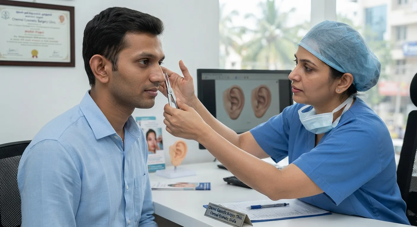
Prominent or protruding ears can significantly impact an individual's self-image, social confidence, and psychological well-being from early childhood through adulthood. For individuals and parents researching otoplasty in Chennai, modern aesthetic and reconstructive ear surgery offers a permanent, safe, and anatomically precise solution to restore natural facial harmony. This guide details everything you need to know about otoplasty, from surgical techniques and age considerations to recovery timelines and local cost factors.
What Is Otoplasty and How Does Prominent Ear Correction Work?
Otoplasty, commonly referred to as ear pinning or cosmetic ear surgery, is a specialized surgical procedure designed to alter the shape, position, proportion, or projection of the auricle (the external ear, also called the pinna). The primary objective of prominent ear correction is to bring protruding ears closer to the sides of the head, recreate missing or poorly defined natural anatomical folds, and establish bilateral symmetry across the facial profile.
The human external ear is composed of a delicate, intricate framework of fibroelastic cartilage covered by thin perichondrium and skin. In a normally positioned ear, the auricle projects from the side of the head at an angle between 15 and 20 degrees, with the distance from the outer helical rim to the scalp measuring roughly 15 to 20 millimeters. When this angle exceeds 20 to 25 degrees, or the helical distance exceeds 20 millimeters, the ear appears overtly prominent.
During an otoplasty, a facial plastic or reconstructive surgeon reshapes, folds, or reduces this structural cartilage framework. By placing specialized internal, non-absorbable retention sutures and selectively thinning or scoring the cartilage, the surgeon secures the pinna in a natural anatomical position without disrupting the internal mechanics of hearing.
Aesthetic vs. Functional Ear Surgery: Understanding the Difference
It is vital to distinguish between aesthetic ear reshaping and functional middle ear surgery. While otoplasty modifies the external cartilage and overlying skin for structural and cosmetic refinement, it does not alter the external auditory canal, the tympanic membrane (eardrum), or the ossicular chain responsible for sound transmission.
Patients suffering from functional middle ear conditions, such as hearing loss or chronic drainage, require otologic procedures rather than aesthetic pinna reshaping. For instance, interventions like tympanoplasty and mastoidectomy address underlying structural infections and perforations within the middle ear cavity, which are entirely separate from cosmetic otoplasty.
Understanding the Causes of Prominent and Protruding Ears
Prominent ears are predominantly congenital, meaning they develop during fetal growth and are present at birth. While the condition rarely causes functional hearing impairment, its anatomical origins stem from specific genetic and structural variations in the auricular cartilage.
Anatomical Factors: Antihelical Fold vs. Conchal Bowl Overgrowth
In clinical practice, prominent ears are generally caused by two distinct anatomical abnormalities, which can occur individually or together:
- Underdeveloped or Absent Antihelical Fold: The antihelix is the Y-shaped internal fold of cartilage that gives the mid-to-upper ear its contoured, curved shape. When the cartilage fails to fold backward during development, the outer rim (helix) protrudes directly lateral to the head.
- Conchal Hypertrophy (Conchal Bowl Overgrowth): The concha is the deep, cup-shaped cartilage bowl located immediately outside the ear canal entrance. If this bowl is abnormally deep or positioned at an excessive angle off the skull, it pushes the entire pinna forward, even if the antihelical fold is normally formed.
In addition to genetic inheritance patterns, acquired prominent ear deformities can result from direct trauma, lacerations, or poorly healed ear hematomas (such as cauliflower ear deformities). A specialized clinical evaluation determines whether the deformity is purely cartilaginous, soft tissue-based, or combined.
Ideal Age for Prominent Ear Correction: When is Surgery Recommended?
One of the most frequent questions regarding prominent ear correction is the optimal age for intervention. The timing of otoplasty is guided by both biological cartilage development and psychosocial considerations.
Pediatric Considerations: Cartilage Pliability and Timing
By age 5 to 6, the human auricle has reached approximately 85% to 90% of its full adult size. At this stage of development:
- The cartilage has developed sufficient structural integrity to hold internal reshaping sutures securely.
- The cartilage remains exceptionally soft, pliable, and elastic, allowing surgeons to shape natural folds with minimal tension.
- Correcting the prominence before entering primary school spares children from playground teasing and the self-consciousness that often emerges in school environments.
During the first 6 to 8 weeks of infancy, non-surgical ear molding or splinting may correct minor deformities due to high levels of maternal estrogen circulating in the infant's bloodstream. However, once maternal hormones decline and the cartilage hardens after the second month of life, surgical otoplasty becomes the only definitive treatment. Parents evaluating other early-childhood ear concerns may also find our guide to pediatric ENT conditions and treatments helpful.
Adult Otoplasty: Aesthetic Goals and Realistic Expectations
There is no upper age limit for prominent ear correction. Many adults who could not undergo the procedure in childhood seek otoplasty later in life to correct lifelong insecurities, improve professional confidence, or wear short hairstyles. In adult patients, the ear cartilage is thicker, firmer, and occasionally calcified. While this requires refined surgical techniques to prevent cartilage fracture or unnatural edges, the aesthetic results in adult otoplasty are equally predictable, highly stable, and permanent.
Key Differences Between Pediatric and Adult Ear Pinning Surgery
Although the fundamental goal—creating a natural, proportional ear contour—remains identical across all age groups, perioperative management varies between children and adults.
| Feature | Pediatric Otoplasty (Ages 5–12) | Adult Otoplasty (Teens & Adults) |
|---|---|---|
| Primary Anaesthesia | General anaesthesia | Local anaesthesia + IV sedation |
| Cartilage Texture | Pliable, soft, easily contoured | Firmer, calcified, springy |
| Surgical Invasiveness | Primarily suture-reshaping | Suture + cartilage thinning/relief |
| Post-Op Compliance | Requires parental monitoring | Self-managed aftercare |
| Recovery Environment | Daycare or overnight pediatric stay | Outpatient / daycare discharge |
- Pediatric Patients: Children generally undergo otoplasty under general anaesthesia administered by a dedicated pediatric anaesthesiologist. This ensures complete immobility, avoids distress during the procedure, and allows the surgical team to perform delicate, symmetrical cartilage sculpting in a controlled environment.
- Adult Patients: Adults typically undergo otoplasty under local anaesthesia accompanied by conscious IV sedation. The local anaesthetic blocks the sensory nerves of the ear, making the procedure completely painless while allowing for a rapid recovery and same-day discharge.
Clarifying the Scope: Why Otoplasty is Structural, Not Hearing Surgery
A frequent misconception among patients is that prominent ear surgery might alter, improve, or inadvertently damage auditory function. It is important to emphasize that otoplasty is strictly external structural surgery. The external ear acts as a funnel to capture sound waves, directing them into the ear canal. Reshaping or repositioning the cartilaginous framework of the pinna does not change the canal diameter or alter auditory acuity.
When to Consult an Otologist vs. a Facial Plastic Surgeon
Before undergoing cosmetic ear surgery, patients must be properly evaluated to ensure their concerns are isolated to the external auricle. Understanding what an ear, nose, and throat doctor is called and the sub-specialties within ENT is helpful:
- Consult an Otologist / Neurotologist when: The patient experiences hearing loss, persistent ringing (tinnitus), vertigo, chronic foul-smelling ear drainage, or suspected middle ear pathology such as an ear drum perforation. Pediatric patients with persistent middle ear fluid may require ear tubes (grommets) rather than aesthetic surgery.
- Consult a Facial Plastic / Reconstructive Surgeon when: The goal is repositioning protruding ears, repairing torn earlobes, reconstructing traumatic avulsions, or rebuilding congenitally malformed auricular frameworks (microtia).
Advanced Auricular Reconstruction: Microtia, Trauma, and Acquired Defects
Beyond standard cosmetic prominent ear pinning, reconstructive ear surgery addresses severe congenital anomalies, extensive physical trauma, and post-oncologic defects.
Microtia Repair and Autologous Cartilage Grafting
Microtia is a congenital condition occurring in roughly 1 in 5,000 to 8,000 births, where the external ear is underdeveloped, rudimentary, or entirely absent (anotia). Reconstructing an ear in microtia requires advanced reconstructive surgery, typically performed in multiple planned stages:
- Stage 1 (Cartilage Framework Harvest & Fabrication): Autologous rib cartilage is harvested from the patient's chest wall. The surgeon meticulously carves and wires these cartilage fragments into a three-dimensional framework replicating the antihelix, helix, tragus, and conchal bowl. This framework is embedded beneath a skin pocket at the ear site.
- Stage 2 (Ear Elevation & Lobule Transposition): Several months later, the reconstructed framework is elevated from the mastoid bone to match the projection of the opposite ear, supported by a cartilage wedge and covered with a skin graft or vascularized fascial flap.
- Synthetic Alternatives: In select cases, porous polyethylene synthetic frameworks covered by a temporoparietal fascia flap provide an alternative for patients where rib harvesting is contraindicated.
Traumatic Ear Reconstruction and Scar Revision
Trauma from motor vehicle collisions, sports injuries, animal bites, burns, or lacerations can cause partial or total avulsion of the ear. Rebuilding the ear after trauma involves debriding non-viable tissue, re-aligning sheared cartilage edges with micro-sutures, and applying local rotation flaps (such as preauricular or postauricular skin flaps) to recreate a natural aesthetic rim.
Split Earlobe and Earlobe Deformity Correction
Earlobe tears and deformities represent one of the most common minor reconstructive procedures performed in clinical practice. Unlike the upper two-thirds of the pinna, the earlobe (lobule) contains no cartilage; it is composed entirely of fibrofatty tissue, skin, and an extensive microvascular network.
In-Office Split Earlobe Repair Procedure
Earlobe defects usually stem from chronic wear of heavy dangling earrings, sudden downward traction, or intentional gauge piercing:
- Surgical Technique: Earlobe repair is performed under local anaesthesia in a minor surgical suite. Because a split piercing track is lined with healed skin, the surgeon carefully excises the inner tract.
- Tissue Re-approximation: The two freshened tissue margins are brought together in multiple layers using fine sutures. To prevent a visible notch at the bottom border of the earlobe, surgeons often employ a miniature Z-plasty or lap-joint flap rather than a straight-line closure.
- Downtime: The procedure takes approximately 20 to 30 minutes per earlobe, involves minimal discomfort, and allows patients to resume normal non-strenuous daily activities immediately.
Guidelines for Safe Re-Piercing After Earlobe Surgery
- Healing Window: The earlobe must heal for at least 6 to 12 weeks to allow collagen maturation and tensile strength restoration.
- Offset Location: The new piercing must never be placed directly through the surgical scar tissue, as scar tissue is weaker than native tissue and will quickly tear again. It should be positioned at least 2 to 3 millimeters away from the scar line.
- Stud Selection: Patients should wear lightweight, hypoallergenic titanium or gold studs for the first 6 months before using heavier earrings.
The Otoplasty Surgical Procedure: Techniques and Incision Placement
Modern otoplasty techniques prioritize long-term stability and a soft, natural contour. Overly aggressive cartilage excision can lead to sharp, unnatural ridges or the characteristic "pinned-back" telephone ear deformity.
Cartilage-Sparing vs. Cartilage-Resection Techniques
- Cartilage-Sparing Techniques (Mustardé and Furnas Suturing):
- Mustardé Technique: The surgeon exposes the posterior cartilage and places horizontal mattress sutures. Tightening these permanent sutures gently folds the cartilage to create a smooth, rounded antihelical fold.
- Furnas Technique: To correct conchal bowl prominence, non-absorbable concha-mastoid sutures anchor the deep conchal cartilage directly back to the fibrous periosteum over the mastoid bone.
- Cartilage-Resection / Cartilage-Cutting Techniques:
- When conchal hypertrophy is severe, a small crescent of excess conchal cartilage is excised to allow the ear to rest closer to the head without excessive suture tension.
- Controlled anterior cartilage scoring exploits cartilage elasticity: scoring one side causes it to naturally curl away from the scored surface, forming a natural contour.
Incision Placement and Hidden Scarring
All primary otoplasty incisions are placed discreetly on the posterior surface of the ear, directly within the retroauricular sulcus (the natural groove between the back of the ear and the side of the head). Once fully healed, the resulting fine scar line is completely hidden from front, side, and rear view. Surgical procedures typically take 1 to 2 hours for both ears. For patients who use tobacco, it is critical to note that smoking causes significant microvascular impairment, delaying wound healing and increasing the risk of cartilage necrosis; smoking cessation is mandated for at least 3 to 4 weeks before and after surgery.
Recovery Timeline, Aftercare, and Long-Term Results
Understanding the postoperative protocol ensures a smooth recovery and preserves the delicate structural repositioning achieved during surgery.
| Recovery Phase | Clinical Focus & Patient Guidelines |
|---|---|
| Days 1 to 3 | Bulky supportive head dressing remains intact; mild ache managed with oral analgesics; head elevated at 30–45 degrees. |
| Days 5 to 7 | Primary dressing removed by surgeon; ears inspected; light elastic headband applied; hair can be gently washed. |
| Weeks 2 to 3 | Non-absorbable skin sutures removed; swelling significantly drops; return to school or desk work permitted. |
| Weeks 4 to 6 | Night-time headband use phased out; non-contact exercise resumed; avoid contact sports. |
| Months 3 to 6 | Residual swelling resolves; cartilage stabilizes; final aesthetic contour established. |
Post-Surgical Headband and Dressing Protocol
Immediately following surgery, a soft, compressive head dressing is applied. This prevents postoperative hematoma formation, minimizes edema, and splints the ears in their new position. After the initial bulky dressing is removed on day 5 to 7, patients transition to a soft, elastic headband. This must be worn 24 hours a day for the first two weeks, and subsequently worn only during sleep for an additional 4 weeks. Night-time protection prevents the ear from inadvertently folding forward against the pillow while turning during sleep.
Managing Swelling, Bruising, and Sleeping Positions
- Sleeping Position: Patients must sleep on their back with their head elevated on two pillows for the first 2 to 3 weeks. Side sleeping is strictly prohibited to avoid asymmetric pressure or suture disruption.
- Bruising and Sensitivity: Moderate bruising and altered skin sensation (numbness or hypersensitivity to cold) are common and resolve over 4 to 8 weeks as cutaneous nerves regenerate.
Otoplasty Cost in Chennai: Key Pricing Factors and Inclusions
The financial investment for prominent ear correction in Chennai varies depending on the clinical complexity of the ear deformity, surgical facility standards, and procedural approach.
| Procedure Type | Typical Cost Range (INR) | Key Inclusions |
|---|---|---|
| Split Earlobe Repair (Bilateral) | ₹8,000 – ₹20,000 | Local anaesthesia, in-office procedure, suture removal |
| Standard Bilateral Otoplasty (Adult / Local + Sedation) | ₹45,000 – ₹95,000 | Surgeon fees, daycare OT charges, post-op dressings, follow-up |
| Pediatric Bilateral Otoplasty (General Anaesthesia) | ₹65,000 – ₹1,25,000 | General anaesthesia, pediatric team, daycare/overnight stay |
| Complex Microtia Reconstruction (per stage) | ₹1,50,000 – ₹3,50,000 | Multi-stage rib graft harvest, inpatient admission |
Costs are indicative estimates and vary with clinical evaluation and hospital infrastructure.
Key Factors Influencing Overall Treatment Expenses
- Surgical Complexity: Bilateral prominent ear pinning involving both conchal setback and antihelical fold recreation requires more surgical time than a unilateral or minor adjustment.
- Anaesthesia Modality: Pediatric cases requiring general anaesthesia incur anaesthesiologist fees and dedicated monitoring that are not required for adult local anaesthesia cases.
- Surgeon Expertise: Reconstructive and facial plastic surgeons specializing in head and neck anatomy provide higher precision, reducing the risk of revision surgery or cartilage irregularities.
- Insurance Coverage: Purely cosmetic otoplasty is considered elective and is generally not covered by health insurance. However, structural reconstruction of congenital deformities (microtia/anotia) or acute trauma repair often qualifies for partial or complete insurance reimbursement.
Expert Facial Plastic and Reconstructive Ear Surgery at THANC Hospital, Chennai
When considering otoplasty, selecting a specialized medical center with dedicated head and neck surgical infrastructure is critical to achieving natural, safe, and balanced results.
Located in Kilpauk, Chennai, THANC Hospital is a leading super-speciality surgical center. Otoplasty is performed by the dedicated Facial Plastic Surgery team of Dr. Vidhyadharan S, Clinical Director at THANC Hospital, with every patient admitted under his care.
Patients undergoing ear correction at THANC Hospital benefit from:
- Tailored Aesthetic Mapping: Comprehensive pre-surgical photographic analysis and anatomical measurements to plan symmetrical, individualized ear projection.
- Advanced Operating Suites: Laminar airflow surgical suites equipped for microvascular, pediatric, and intricate cartilage-shaping interventions.
- Multidisciplinary Collaborative Care: On-site collaboration between facial plastic surgeons, pediatric anaesthesiologists, and ENT sub-specialists ensures comprehensive safety for both elective cosmetic patients and complex pediatric reconstructive cases.
Whether correcting prominent ears in a school-aged child or refining external ear contours for an adult, THANC Hospital provides compassionate, clinically rigorous care in the heart of Chennai.
Frequently Asked Questions
Will otoplasty leave visible scars on my ears?
No. The surgical incisions for otoplasty are positioned within the retroauricular sulcus—the natural fold behind the ear. Once healed, the scar is concealed behind the auricle and is not visible from the front, side, or rear.
Is prominent ear correction painful?
During surgery, patients feel no pain due to general or local anaesthesia. Postoperatively, patients experience mild to moderate aching or pressure around the ears for the first 48 to 72 hours, which is effectively controlled with standard oral pain medications.
Can prominent ears return to their original protruding position after surgery?
When performed using proper structural cartilage-sparing techniques and permanent internal retention sutures, otoplasty results are permanent. Relapse occurs in a small minority of cases, typically associated with premature trauma or failure to wear the protective night headband during early recovery.
How soon can I or my child return to work or school after otoplasty?
Most pediatric and adult patients return to school or desk work within 7 to 10 days, following the removal of the primary compressive head dressing. However, contact sports, strenuous workouts, swimming, and helmet use must be avoided for 4 to 6 weeks.
Does otoplasty affect my ability to hear?
No. Otoplasty reshapes only the external cartilaginous auricle. It does not alter the ear canal, eardrum, middle ear cavity, or internal auditory nerve pathways. Hearing remains completely unaffected.
Learn more about Facial Plastic Surgery
View treatments, doctors & FAQs
Concerned about this condition?
Our specialists can help. Book a consultation today.
Book Consultation
MBBS, MS (ENT), DNB (ENT), MCh (Head & Neck), FICRS, FEB – ORL HNS, ASOHNS
Clinical Director & Senior Consultant Surgeon