In this article
- What is an Alveolar Bone Graft?
- Who Needs an Alveolar Bone Graft?
- How the Alveolar Bone Graft is Performed
- Preparing for an Alveolar Bone Graft
- Recovery After an Alveolar Bone Graft
- Risks and Complications
- Results and Success Rate
- Why Choose THANC Hospital for an Alveolar Bone Graft?
- Frequently Asked Questions
What is an Alveolar Bone Graft?
An alveolar bone graft is a highly specialized surgical procedure designed to repair a gap in the upper jawbone. The alveolus is the specific part of the jawbone that holds the roots of your teeth in place. When a child is born with a cleft lip and palate, this bony ridge often fails to fuse together during pregnancy. This leaves a distinct notch or a complete gap in the gum line.
During this surgery, the oral and maxillofacial surgeon takes healthy bone from another part of the body and places it into this gap. This new bone acts as a bridge, connecting the two separated sides of the upper jaw. Over time, the transplanted bone heals and fuses with the existing jawbone, creating a single, continuous, and solid dental arch.
Doctors perform a bone graft cleft palate procedure to solve several critical functional and structural problems. First and foremost, it provides a solid foundation of bone for the permanent teeth to grow into. Without this bone, teeth near the cleft will not erupt properly or may be lost entirely.
Secondly, this procedure closes any remaining oronasal fistula. A fistula is an abnormal hole that connects the roof of the mouth directly to the nasal cavity. Closing this hole prevents food and liquids from passing from the mouth into the nose during meals. It also helps improve speech by preventing air from escaping through the nose when talking.
Finally, the bone graft provides essential structural support for the base of the nose. Children with a cleft often have a flattened or asymmetrical nasal appearance on the side of the cleft. By building up the bone underneath the nose, the surgeon helps lift and support the nasal cartilage. This significantly improves facial symmetry and prepares the foundation for any future cosmetic adjustments to the nose.
Who Needs an Alveolar Bone Graft?
Any child born with a complete cleft of the gum ridge will eventually require this procedure. While the initial surgeries to close the lip and the roof of the mouth are performed during infancy, the jawbone itself requires attention later in childhood. Conservative treatments, such as braces alone, cannot fix this problem. Bone does not naturally grow across an empty space, and teeth cannot be moved into an area where no bone exists to support them.
The timing of this surgery is incredibly important. The standard and most successful approach is called a secondary alveolar bone graft. Surgeons typically perform this procedure when the child is between 8 and 11 years old. The exact timing depends entirely on the child's dental development rather than their chronological age.
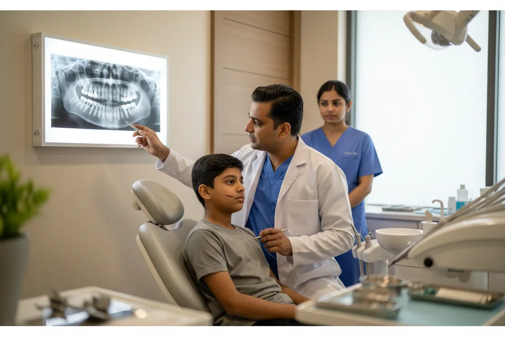
The surgeon must place the new bone before the permanent maxillary canine (the upper eye tooth) erupts into the mouth. Dental X-rays will show when the root of this canine tooth is about half to two-thirds formed. This is the perfect window for surgery. Placing the graft at this exact stage encourages the canine tooth to erupt naturally through the newly placed bone, which helps keep the graft strong and healthy for a lifetime.
In India, roughly 1 to 1.5 in every 1,000 children are born with a cleft lip or palate. This makes cleft conditions one of the most common birth defects in the country. Many families successfully complete the primary lip and palate repairs when their baby is young. However, due to a lack of awareness, some families miss the important 8-to-11-year-old window for the bone graft.
Ideal candidates for this surgery are children who have maintained excellent oral hygiene and have completed their preliminary orthodontic treatment. If a patient misses this childhood window, surgeons can still perform a late bone graft in adulthood. However, adult patients may require dental implants later because the natural teeth near the cleft may have already been lost or impacted.
How the Alveolar Bone Graft is Performed
The alveolar bone graft is a complex inpatient surgery performed in a hospital operating room. The entire procedure takes about two to three hours to complete. The patient receives general anesthesia, meaning they will be completely asleep, unaware of the surgery, and feeling no pain throughout the process.
The surgery involves two main phases: harvesting the bone and placing the graft. The gold standard for this procedure is using the patient's own bone, known as an autograft. The surgeon typically harvests this bone from the iliac crest, which is the thick edge of the hip bone.
To harvest the bone, the surgeon makes a small incision just below the waistline. They carefully open the hard outer shell of the hip bone to access the cancellous bone inside. Cancellous bone is soft, spongy, and rich in live bone-forming cells and stem cells. The surgeon scoops out the necessary amount of this spongy bone and then closes the hip incision, leaving the structural integrity of the hip completely intact.
Next, the surgical team focuses on the mouth. The surgeon makes precise incisions in the gum tissue surrounding the cleft gap. They carefully separate the tissue that lines the nasal cavity from the tissue that lines the mouth. The surgeon then stitches the nasal lining closed, creating a watertight ceiling above the gap. This step is important to separate the mouth from the nose permanently.
Once the area is prepared and cleaned, the surgeon takes the spongy bone harvested from the hip and packs it tightly into the cleft defect. They ensure the bone fills the entire gap from the base of the nose down to the gum line. Finally, the surgeon stretches the gum tissue over the new bone graft and stitches it securely in place. These stitches are usually dissolvable and will fall out on their own over a few weeks.
After the surgery, the patient is moved to a recovery room to wake up from the anesthesia. Most patients will need to stay in the hospital for one to three days for observation and pain management. You can learn more about the surgical expertise required for this procedure on our Oral & Maxillofacial Surgery speciality page.
Preparing for an Alveolar Bone Graft
Preparation for a bone graft cleft palate surgery begins several months before the actual operation. The most critical part of this preparation involves working closely with an orthodontist. Children with a cleft often have an upper jaw that is narrow or collapsed inward on the side of the cleft.
To fix this, the orthodontist will place a custom device called a palatal expander inside the child's mouth. This device gently widens the upper jaw over several months. Expanding the jaw creates enough physical space for the surgeon to access the cleft and place the bone graft properly. It also aligns the two segments of the upper jaw into a proper arch shape.
As the surgery date approaches, the surgical team will order several pre-operative tests. These include standard blood tests to ensure the patient is healthy enough for general anesthesia. The surgeon will also take advanced 3D X-rays, such as a Cone Beam Computed Tomography (CBCT) scan. This scan provides a highly detailed, three-dimensional view of the jawbone, the exact size of the cleft, and the precise location of the unerupted canine tooth.
Excellent oral hygiene is absolutely mandatory before surgery. The mouth is naturally full of bacteria, and any existing plaque or gum inflammation can severely increase the risk of the new bone graft getting infected. The patient must brush and floss diligently. The dental team will perform a professional cleaning a few days before the surgery and may prescribe an antibacterial mouthwash.
You will receive specific instructions on what medications to stop taking before the procedure. You must stop any herbal supplements, vitamins, or blood-thinning medications at least two weeks prior, as these can increase bleeding during surgery.
On the day of the surgery, bring loose, comfortable clothing for the patient. Button-down shirts are highly recommended because they do not need to be pulled over the face and head, which could disturb the surgical site. Parents should also prepare their home in advance by stocking the kitchen with appropriate soft foods and liquids. For more information on preparing for major jaw procedures, you can read our guide on Orthognathic Surgery Recovery Week by Week.
Recovery After an Alveolar Bone Graft
The recovery period requires patience and strict adherence to the surgeon's guidelines. During the first 24 to 48 hours, swelling in the upper lip, cheeks, and nose is completely normal. The patient will also experience soreness in the hip area where the bone was harvested. Walking will be difficult and uncomfortable at first, but the hospital staff will provide intravenous (IV) pain medication to keep the patient comfortable.
During the first week at home, rest is the primary focus. The patient may walk with a slight limp due to the hip incision. The diet during this first week must be strictly liquid. The mouth will be very sensitive, and chewing is strictly forbidden. Patients should consume nutrient-rich liquids like milkshakes, strained soups, and nutritional supplement drinks.
By the second and third weeks, the facial swelling will significantly decrease. The hip pain will improve, allowing the patient to walk more normally. Most children can return to school during this time, provided they avoid any strenuous physical activities. The diet can now progress to very soft, non-chew foods.
For Indian patients, a soft diet can include well-mashed idli soaked in mild sambar, plain khichdi, curd rice, soft upma, and dal soup. You must avoid anything spicy, crunchy, sticky, or hard. Foods like murukku, raw carrots, nuts, and tough meats are strictly prohibited. These hard foods can easily break the stitches or put pressure on the healing gums, causing the graft to fail.
Weeks four through six mark the critical phase of bone healing. The spongy bone from the hip begins to establish a new blood supply and fuse with the surrounding jawbone. The hip pain usually resolves completely by this stage. However, activity restrictions remain in place.
Patients must avoid all contact sports, such as cricket, football, basketball, or kabaddi, for at least six to eight weeks. A sudden blow to the face could dislodge the healing bone graft. A fall could also injure the healing hip bone.
Oral hygiene during recovery requires special care. The patient cannot use a toothbrush directly on the surgical site until the surgeon gives permission. Instead, they must gently rinse their mouth with a prescribed antibacterial mouthwash or warm salt water after every single meal. Keeping the surgical area free of food particles is vital for preventing infection. To understand more about recovering from jaw-related procedures, explore our article on Corrective Jaw Surgery Types.
Risks and Complications
Like any major surgical procedure, an alveolar bone graft carries certain risks. The surgical team takes extensive precautions to minimize these issues, but patients and parents must be aware of what to expect. Common, minor risks include temporary swelling, bruising on the face and hip, and mild bleeding from the nose or mouth during the first few days. Hip pain is also an expected part of the healing process and is managed with oral pain relievers.
However, there are rare but serious risks associated with this surgery. The most significant complication is graft failure. This happens when the transplanted bone does not survive in its new location. Graft failure is usually caused by an infection, poor blood supply to the area, or excessive pressure on the gums from eating hard foods too soon. If the graft fails, the body will absorb the dead bone, and a second surgery will be required months later.
Another risk is wound breakdown, where the stitches holding the gums together open up prematurely. This exposes the underlying bone graft to the bacteria in the mouth, significantly increasing the risk of infection and failure. Damage to the roots of adjacent teeth is also a rare possibility during the preparation of the cleft site.
THANC Hospital minimizes these risks through meticulous surgical planning and execution. Our surgical team uses advanced 3D imaging to map the exact anatomy of the cleft and the surrounding tooth roots before making a single incision. We follow strict sterilization protocols to prevent infection. Furthermore, our surgeons use precise tissue-handling techniques to ensure the gums are closed securely without any tension, drastically reducing the risk of wound breakdown.
Results and Success Rate
When performed at the correct time and by an experienced surgical team, the alveolar bone graft has an exceptionally high success rate of over 90%. The primary outcome is the creation of a continuous, solid upper jawbone. This structural integrity is essential for the long-term health of the patient's mouth.
A successful graft allows the permanent canine tooth to erupt naturally through the new bone. Once this tooth comes in, it stimulates the grafted bone, keeping it strong and dense. The orthodontist can then use braces to move this tooth into its perfect position, completing the dental arch. If a patient is missing a tooth entirely, the new bone provides the necessary foundation for a dental implant later in life.
The results of this surgery are permanent. Over several months, the body remodels the spongy hip bone, turning it into dense jawbone. Eventually, the grafted bone becomes completely indistinguishable from the natural bone surrounding it.
Beyond dental health, the surgery provides significant aesthetic and functional benefits. The closure of the oronasal fistula improves eating and speaking. The added bone volume supports the base of the nose, lifting the nasal cartilage and improving facial symmetry. This thorough improvement in both function and appearance greatly enhances the patient's quality of life. For more information on how jaw surgeries improve facial structure, read our guide on Underbite Overbite Surgical Non-Surgical Correction.
Why Choose THANC Hospital for an Alveolar Bone Graft?
THANC Hospital offers a team-based approach to cleft lip and palate care. Dr. M. Veerabahu has decades of specialized experience in complex oral and maxillofacial surgeries, supporting the highest standard of precision and safety. Our team coordinates smoothly with orthodontists, speech therapists, and pediatric specialists to guide your child through every phase of their treatment journey. We are dedicated to providing compassionate care and achieving the best possible functional and aesthetic outcomes for our patients. To learn more about our lead surgeon, visit the profile of Dr. M. Veerabahu, or Book an Appointment to discuss your treatment options.
Frequently Asked Questions
Does the hip bone grow back after the bone graft cleft palate surgery?
Yes, the hip bone does heal and regenerate. The surgeon only removes the soft, spongy bone from the inside of the hip, leaving the hard outer shell completely intact. Your body will naturally fill in this hollowed-out area with new bone tissue over the course of a few months.
Will my child be in a lot of pain after the procedure?
Your child will experience some discomfort, particularly in the hip area when trying to walk or move. However, the surgical team will provide effective intravenous pain medication while in the hospital and prescribe oral pain relievers for home use. Most children find the pain very manageable after the first few days.
Can we use artificial bone instead of taking bone from the hip?
While artificial bone substitutes and donor bone do exist, using the patient's own hip bone remains the absolute gold standard. The hip bone contains live bone-forming cells and proteins that offer the highest success rate for integrating into the jaw. Artificial bone does not have these live cells and has a higher rate of failure in cleft repairs.
What happens if we delay the alveolar bone graft surgery?
Timing is critical for this procedure. If you delay the surgery until after the permanent canine tooth tries to erupt, the tooth will not have any bone to support it. This can lead to the permanent loss of the tooth and make future orthodontic correction much more difficult.
How long will my child need to eat a soft diet?
Your child will need to adhere to a strict liquid and very soft food diet for about four to six weeks. Eating hard, crunchy, or chewy foods too early can tear the stitches or put pressure on the healing gums. Protecting the surgical site from chewing forces is essential for the bone graft to survive.
When can my child start playing sports again?
Your child must avoid all contact sports, such as cricket, football, or martial arts, for at least six to eight weeks after surgery. A sudden impact to the face could dislodge the healing bone graft in the jaw. Additionally, a fall could injure the hip bone while it is still recovering from the harvest procedure.
Learn more about Oral & Maxillofacial Surgery
View treatments, doctors & FAQs
Concerned about this condition?
Our specialists can help. Book a consultation today.
Book Consultation