In this article

What is a Skull Base Tumor?
A skull base tumor is an abnormal growth of cells that develops at the bottom of the skull. The skull base forms the floor of the cranial cavity, acting as a bony shelf that supports your brain. It separates your brain from your facial structures, sinuses, eyes, and ears. This area is highly complex and crowded with major blood vessels and cranial nerves (the nerves that control your facial movements, vision, hearing, and smell).
When a tumor grows in this tight space, it can press against these critical structures. This pressure causes various symptoms depending on the exact location of the growth. A skull base tumor can be either benign (non-cancerous) or malignant (cancerous). Benign tumors grow slowly and do not spread to other parts of the body, but they can still cause severe damage by compressing the brain and nerves. Malignant tumors grow aggressively and can invade surrounding bone and tissue.
Skull base tumors can affect people of all ages, though certain types are more common in specific age groups. In India, these tumors present a significant health challenge. Pituitary adenomas, which are the most common type of skull base tumor, account for 10 to 15 percent of all brain tumors. Medical registries report that more than one million cases of pituitary adenomas occur in India every year.
Another specific type of skull base tumor, nasopharyngeal carcinoma, shows a unique geographic pattern in India. While it remains rare in most parts of the country, it is highly prevalent in the Northeastern states. For example, districts in Nagaland report an incidence rate of up to 19.4 cases per 100,000 people, which is among the highest rates globally.
Common Types of Skull Base Tumors
Doctors classify skull base tumors based on where they start and the type of cells involved. The most common types include:
- Pituitary Adenomas: These benign growths develop in the pituitary gland, a small organ located centrally at the skull base. They can disrupt normal hormone production.
- Meningiomas: These tumors grow from the meninges (the protective membranes surrounding the brain and spinal cord). Most meningiomas are benign and grow very slowly.
- Acoustic Neuromas (Vestibular Schwannomas): These benign tumors develop on the nerve that connects the ear to the brain, directly affecting hearing and balance.
- Chordomas and Chondrosarcomas: These are rare, slow-growing malignant tumors that develop from the bone and cartilage at the base of the skull.
- Craniopharyngiomas: These benign tumors grow near the pituitary gland and often affect vision and hormone levels.
- Nasopharyngeal Carcinomas: This malignant cancer starts in the upper part of the throat behind the nose and can invade the skull base.
Causes and Risk Factors
Researchers do not always know exactly why a skull base tumor develops. Like all tumors, they begin when the DNA inside a cell mutates. These genetic changes instruct the cells to grow and multiply rapidly instead of dying at their natural time. The accumulating cells form a mass or tumor. While the exact trigger for these genetic mutations remains unclear, doctors have identified several factors that increase your risk.
General Causes and Genetic Factors
Some skull base tumors occur due to inherited genetic conditions. If you have a family history of certain genetic syndromes, your risk of developing these tumors increases.
- Neurofibromatosis type 2 (NF2): This genetic disorder greatly increases the risk of developing acoustic neuromas on both hearing nerves.
- Multiple Endocrine Neoplasia type 1 (MEN1): This inherited condition causes tumors to grow in the endocrine glands, significantly increasing the risk of pituitary adenomas.
- Previous Radiation Therapy: If you received radiation treatment to your head or neck for a previous medical condition, you have a higher risk of developing a skull base tumor, such as a meningioma, later in life.
India-Specific Risk Factors
Environmental and lifestyle factors play a major role in the development of certain skull base and head and neck tumors in India.
- Tobacco Use: Smoking cigarettes, bidis, and chewing smokeless tobacco (like gutka or khaini) strongly increase the risk of malignant tumors in the nasal cavity and nasopharynx. Tobacco contains harmful chemicals that damage the cellular DNA in your respiratory tract.
- Dietary Habits: In Northeast India, the traditional diet includes high amounts of smoked meats, fermented foods, and salt-cured fish. Regular consumption of these foods exposes the body to nitrosamines (chemical compounds linked to cancer), which significantly increases the risk of nasopharyngeal carcinoma.
- Viral Infections: Infection with the Epstein-Barr Virus (EBV) is a major risk factor for nasopharyngeal carcinoma. This virus is common worldwide, but it interacts with genetic and dietary factors in certain Indian populations to trigger tumor growth.
- Occupational Exposures: Many people in India work in industries that expose them to harmful airborne particles. Inhaling wood dust, leather dust, formaldehyde, and heavy industrial chemicals over many years increases the risk of developing malignant tumors in the nasal cavity and anterior skull base.

Signs and Symptoms
Because the skull base is a crowded area, a growing tumor will eventually press on the brain, blood vessels, or cranial nerves. The symptoms you experience depend entirely on the size, type, and exact location of the tumor. Many benign skull base tumors grow so slowly that they do not cause any symptoms for years.
Early Warning Signs
Patients often mistake the early signs of a skull base tumor for common, less serious conditions like sinus infections or tension headaches. You might notice:
- Persistent headaches that do not improve with standard pain medication.
- A feeling of pressure or fullness in your face and sinuses.
- Chronic nasal congestion or a blocked nose that only affects one side.
- Frequent nosebleeds without an obvious cause.
- A gradual loss of your sense of smell.
Progressive and Serious Symptoms
As the skull base tumor grows larger, it begins to compress the cranial nerves. This compression leads to more noticeable and severe neurological symptoms.
- Visual Changes: You may experience double vision, blurred vision, or a gradual loss of peripheral vision. This happens when the tumor presses against the optic nerves.
- Hearing and Balance Issues: Tumors near the ear can cause tinnitus (ringing in the ears), gradual hearing loss in one ear, dizziness, or a feeling of unsteadiness.
- Facial Nerve Problems: You might feel numbness, tingling, or pain in your face. Some patients develop facial weakness or paralysis on one side.
- Difficulty Swallowing: Tumors pressing on the lower cranial nerves can make it hard to swallow food or change the sound of your voice.
- Clear Fluid Leaking: If a tumor erodes the bone of the skull base, the fluid that surrounds your brain can leak out. You might notice a constant, clear, watery discharge dripping from your nose. You can learn more about this specific symptom in our guide on CSF leak and clear fluid dripping from the nose.
See a Doctor If...
You should schedule an appointment with a specialist if you experience persistent headaches combined with vision changes, unexplained hearing loss in one ear, or continuous clear fluid dripping from your nose. Early detection provides the best chance for successful treatment.

How is a Skull Base Tumor Diagnosed?
Diagnosing a skull base tumor requires a thorough approach. Because the symptoms often mimic other conditions, your medical team will use a combination of physical examinations, specialized imaging, and laboratory tests to confirm the diagnosis.
Clinical and Neurological Examination
Your doctor will start by taking a detailed medical history and asking about your symptoms, lifestyle, and family history. Next, they will perform a thorough neurological examination. The doctor will check your vision, hearing, balance, facial sensation, and muscle strength. This helps the medical team identify exactly which cranial nerves the tumor is affecting.
The doctor may also perform a nasal endoscopy. During this office procedure, the doctor inserts a thin, flexible tube with a light and camera into your nasal passages. This allows the doctor to look directly at the back of your nose and the anterior skull base to check for any visible growths or abnormalities.
Advanced Imaging and Tests
If the clinical exam suggests a problem, your doctor will order advanced imaging tests to get a clear picture of your skull base.
- Magnetic Resonance Imaging (MRI): An MRI uses strong magnets and radio waves to create highly detailed images of your brain, nerves, and soft tissues. This is the best test for identifying a skull base tumor. It shows the exact size, shape, and location of the mass.
- Computed Tomography (CT) Scan: A CT scan uses X-rays to create detailed cross-sectional images of your bones. Doctors use CT scans to see if the tumor has invaded or destroyed the bony structures of the skull base.
- Endocrine Testing: If your doctor suspects a pituitary tumor, they will order specific blood and urine tests. These tests measure your hormone levels to see if the tumor is producing excess hormones or suppressing normal gland function. You can read more about this process in our detailed post on pituitary tumor symptoms, diagnosis, and treatment.
- Biopsy: In some cases, the doctor needs a tissue sample to determine if the tumor is benign or malignant. The surgeon can often obtain this small sample through the nose using endoscopic tools. A pathologist then examines the cells under a microscope to confirm the exact type of tumor.
Treatment Options
The treatment for a skull base tumor depends on several factors, including the tumor's type, size, location, and whether it is benign or malignant. Your age and overall health also play a important role in the decision-making process. At THANC Hospital, a multidisciplinary team of neurosurgeons, head and neck surgeons, and radiation oncologists work together to create a treatment plan for you.
Observation and Medical Management
If you have a small, benign tumor that does not cause any symptoms, your doctor might recommend observation. This approach, often called "watchful waiting," involves scheduling regular MRI scans to monitor the tumor for any signs of growth.
For certain functioning pituitary tumors, doctors use medical therapy as the first line of treatment. Specific prescription medications can block the tumor from producing excess hormones and may even shrink the tumor over time. If medications successfully control the tumor, you may not need surgery.
Surgical Options
When a tumor grows rapidly, causes severe symptoms, or threatens your vision and brain function, surgery becomes necessary. The primary goal of surgery is to remove as much of the tumor as safely possible without damaging the surrounding nerves and blood vessels.
Historically, surgeons relied on open skull base surgery. This traditional approach requires making an incision in the face or skull and removing a piece of bone to access the tumor. While open surgery remains necessary for certain large or complex tumors, modern medicine offers less invasive alternatives.
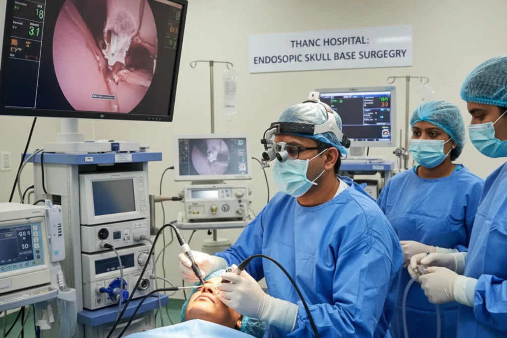
Endoscopic Skull Base Surgery
Today, surgeons treat many of these conditions using endoscopic skull base surgery. This minimally invasive technique allows the surgeon to remove tumors through the natural openings of your nose and sinuses.
During an endoscopic skull base surgery, an ear, nose, and throat (ENT) surgeon and a neurosurgeon work together. They insert an endoscope (a thin, lighted tube with a high-definition camera) into your nostril. The camera projects a magnified view of the skull base onto a large monitor in the operating room.
Using specially designed, long-handled instruments, the surgeons carefully remove the tumor through the nasal passages. This approach offers several major benefits for the patient:
- It requires no external incisions on the face or scalp.
- It avoids the need to retract or move the brain, reducing the risk of neurological damage.
- It generally results in less pain and a faster recovery time compared to open surgery.
- It leaves no visible scars.
You can learn more about our surgical capabilities on our Skull Base Surgery specialty page.
Radiation Therapy
If a tumor is malignant, or if the surgeon cannot safely remove the entire benign tumor, your doctor may recommend radiation therapy. Radiation uses high-energy beams to destroy the remaining tumor cells and stop them from growing.
Doctors often use advanced radiation techniques, such as Stereotactic Radiosurgery (like Gamma Knife) or Proton Beam Therapy. These methods deliver highly focused radiation directly to the tumor while sparing the healthy brain tissue nearby.
Living with a Skull Base Tumor / Recovery and Outlook
Recovering from skull base treatment requires time, patience, and careful follow-up. Your exact recovery timeline will depend on the type of treatment you received and your overall health.
What to Expect After Surgery
If you undergo endoscopic skull base surgery, you will likely spend a few days in the hospital. The medical team will monitor your neurological function closely in the intensive care unit for the first 24 hours. You may experience nasal congestion, mild headaches, and fatigue during the first week.
Your doctor will give you specific instructions to protect the surgical site while it heals. You must avoid blowing your nose, bending over, or lifting heavy objects for several weeks. These activities increase the pressure inside your head and can disrupt the healing process or cause a cerebrospinal fluid leak. The doctor will also prescribe nasal saline sprays to keep your nasal passages moist and help clear away crusting.
Long-Term Follow-Up Care
Because skull base tumors can sometimes grow back, long-term follow-up care is essential. You will need to visit your medical team regularly for physical examinations and follow-up MRI scans. If you had a pituitary tumor, you will also need regular blood tests to monitor your hormone levels. Some patients require hormone replacement medication for the rest of their lives if the pituitary gland does not recover its normal function.
Lifestyle Modifications
Adopting a healthy lifestyle can support your recovery and improve your overall quality of life.
- Quit Tobacco: If you use tobacco in any form, quitting is a key step you can take. Tobacco use delays healing and increases the risk of tumor recurrence.
- Eat a Balanced Diet: Focus on a diet rich in fresh fruits, vegetables, and whole grains. Limit your intake of heavily processed foods, smoked meats, and salt-cured items.
- Manage Stress: Dealing with a tumor diagnosis takes a toll on your mental health. Engage in stress-reducing activities like yoga, meditation, or gentle walking.
- Attend Rehabilitation: If the tumor affected your speech, swallowing, or balance, your doctor will refer you to physical or speech therapists to help you regain those functions.
Why Choose THANC Hospital for Skull Base Tumors?
Treating tumors in this delicate area requires a highly coordinated team of specialists. At THANC Hospital, Dr. Vidhyadharan S leads a multidisciplinary team dedicated to providing precise, patient-centered care. We use advanced endoscopic techniques and full treatment planning to ensure you achieve the best possible outcome with the fastest recovery.
If you are experiencing symptoms or need a second opinion, please Book an Appointment with our specialists today.
Frequently Asked Questions
What is the survival rate for a skull base tumor?
The survival rate depends entirely on whether the tumor is benign or malignant, as well as its specific type and location. Benign tumors, like most meningiomas and pituitary adenomas, have excellent long-term survival rates and are often completely curable. Malignant tumors require more aggressive treatment, but early detection significantly improves the overall outlook and survival chances.
Is endoscopic skull base surgery painful?
Most patients experience surprisingly little pain after an endoscopic procedure compared to traditional open surgery. You will likely feel nasal congestion, sinus pressure, and a mild to moderate headache for the first few days. Your medical team will provide effective pain medication to keep you comfortable during your recovery.
How long does it take to recover from skull base surgery?
Recovery times vary based on the complexity of the surgery and your overall health. Most patients who undergo endoscopic surgery can return to light activities and desk work within three to four weeks. However, full healing of the skull base and nasal tissues can take several months, during which you must avoid strenuous physical exertion.
Can a skull base tumor grow back after removal?
Yes, there is always a small risk that a tumor can grow back, even if the surgeon removed all visible parts of it. This happens if microscopic tumor cells remain in the surrounding bone or tissue. Because of this risk, your doctor will schedule regular follow-up MRI scans to monitor the area for many years after your treatment.
Are skull base tumors hereditary?
Most skull base tumors occur randomly and do not run in families. However, a small percentage of these tumors link directly to inherited genetic syndromes. Conditions like Neurofibromatosis type 2 (NF2) and Multiple Endocrine Neoplasia type 1 (MEN1) significantly increase the risk of developing specific tumors in the skull base.
Will I lose my sense of smell after endoscopic surgery?
You may experience a temporary loss or reduction in your sense of smell immediately after surgery due to swelling and nasal packing. For most patients, the sense of smell gradually returns to normal as the nasal passages heal over a few weeks or months. Permanent loss of smell is rare unless the tumor directly involved the olfactory nerves.
Learn more about Skull Base Surgery
View treatments, doctors & FAQs
Concerned about this condition?
Our specialists can help. Book a consultation today.
Book Consultation